



no.45
[Case Studies] Case 5. Incisional Ptosis Correction + Epicanthoplasty: Before/After and Recovery (feat. Scar Tissue)
2026-05-04
This time, let's look at a patient who had incisional ptosis correction together with epicanthoplasty,
see what changed,
and follow how the scars and swelling progressed.
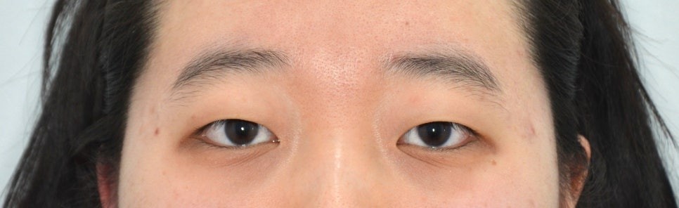
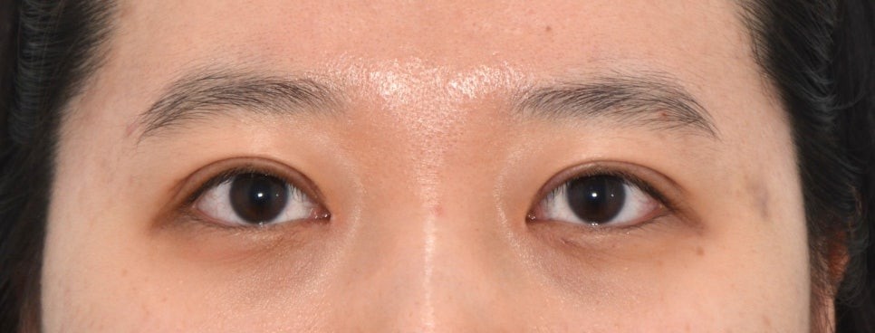
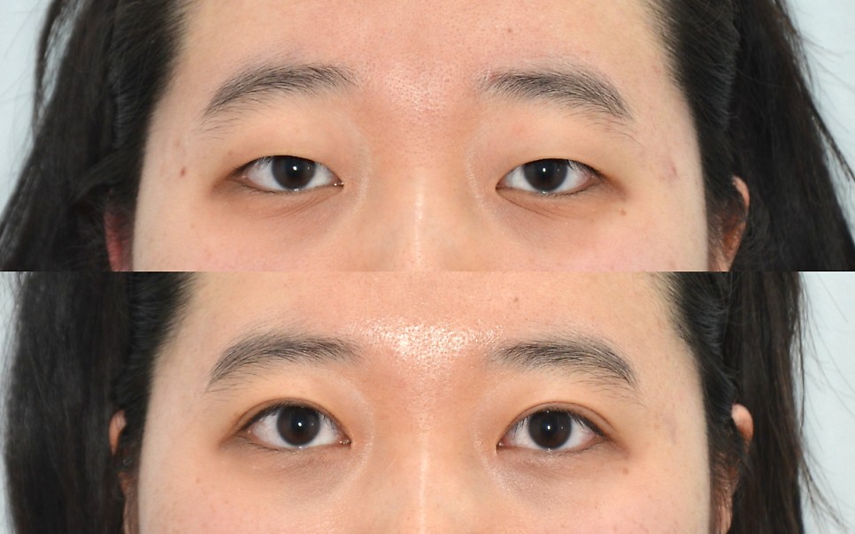
Before surgery,
the dark part of the eye (the iris) was covered and less visible,
giving the eyes a heavy, confined look,
and you can see that she raised her eyebrows to open her eyes.
She was also using the << (left in the photo) eyebrow more.
A fairly prominent epicanthal fold is visible at the inner corners,
along with a long crease extending downward.
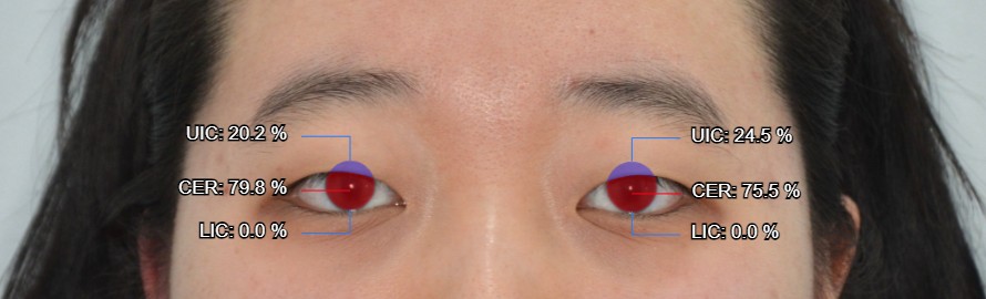
Analysis with AI software
showed that about 75–80% of the iris was exposed.
At least 85% is needed for the eyes not to look heavy,
and around 90% exposure gives a clear, bright look.
Her surgical plan was
incisional double eyelid + ptosis correction
and epicanthoplasty.
This was because there was a lot of loose skin that needed to be removed, her eye-opening strength was weak,
and the epicanthal fold at the inner corners was severe and needed improvement.
This is the pre-operative design.
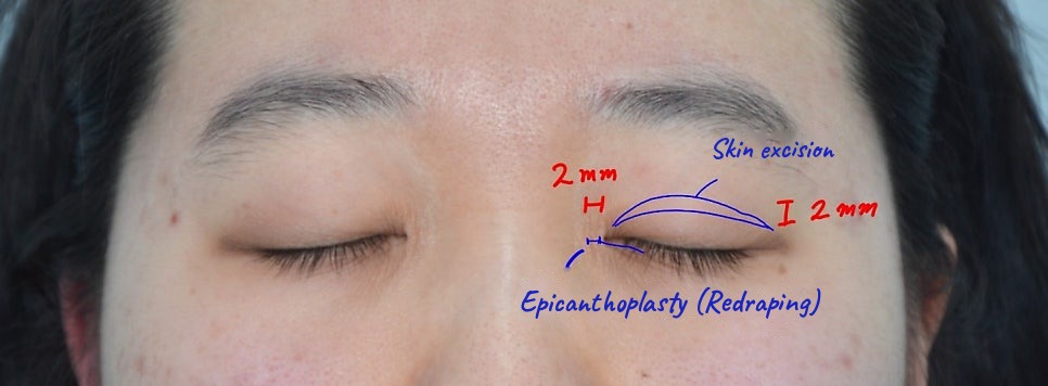
About 2 mm of skin was excised on each side,
and the epicanthoplasty was also about 2 mm on each side.
For ptosis correction, on both sides equally,
a 4.5 mm levator advancement was planned.
1 week after surgery: after stitch removal
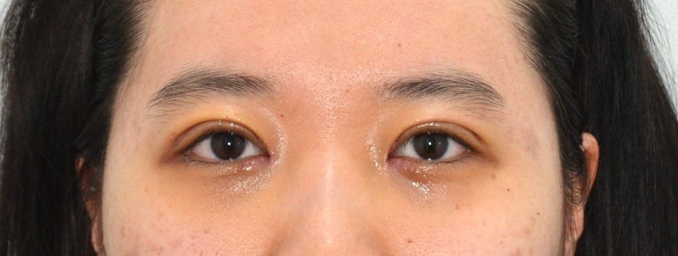
There was less swelling than expected, with slight bruising.
The eyes look much clearer than before surgery,
and you can see that the crease below was reduced by the epicanthoplasty.
1 month after surgery
There was still some swelling,
and in the left eye in the photo, at the inner and outer corners,
the line appeared doubled.
At the inner corner, this was due to slight scarring,
so I gave a scar injection.
The outer corner is something that gradually improves over time.
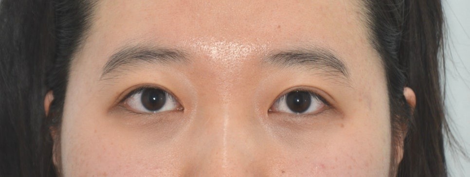
This is 3 months after surgery.
Almost all of the swelling has gone down,
and the issues seen at 1 month have all improved.
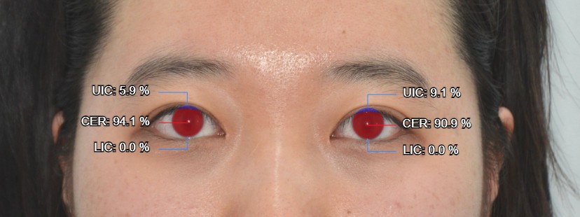
After surgery, iris exposure increased to over 90%,
giving the eyes a clear, bright look.
And the deep crease that started from the inner corner
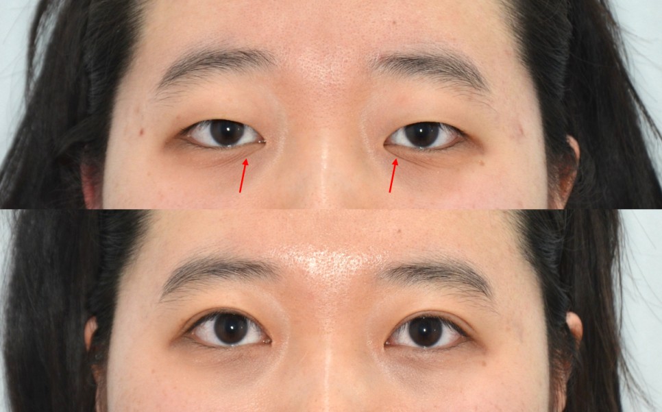
also improved with the epicanthoplasty.
The epicanthoplasty scar is barely visible,
and I expect it to improve further.
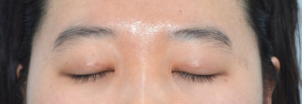
This is how visible the incisional double eyelid scar is.
It is still slightly visible at 3 months, but it will fade further and become invisible over time.
Incisional double eyelid surgery and epicanthoplasty both involve incisions and sutures,
so many people are afraid of scarring and of the recovery,
which is why I wrote this post.
Before / 3 months after
Incisional ptosis correction / Epicanthoplasty
Want to learn more about incisional ptosis correction?
▶ Incisional Ptosis Correction: Technique, Principle, Pros and Cons
Want to learn more about epicanthoplasty?
▶ What Is Inner Corner Eye Surgery (Epicanthoplasty)?
◀ Previous post: Case 4. Non-Incision Double Eyelid Surgery: Before/After and Recovery
Related posts
▶ How to Minimize Scarring After Double Eyelid or Epicanthoplasty Surgery
▶ Who Needs Ptosis Correction (Eye-Opening Surgery)?
WhatsApp Inquiries
https://api.whatsapp.com/message/DE3GWE4FQGMDF1?autoload=1&app_absent=0
Sai Plastic Surgery — Phone Inquiries
Main Line: +82-2-512-4647
* This post was written by Dr. Kwang-rae Kang, Director of Sai Plastic Surgery, with his own text and illustrations, for the purpose of providing medical information in compliance with Article 56(1) of the Korean Medical Service Act.