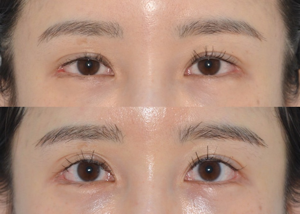
 no.53 [Case Studies] Case 9. Partial Lateral Canthoplasty Reversal and Lower Canthoplasty Revision This time, I'd like to introduce a somewhat unusual case. She had lateral and lower canthoplasty 10 years ago, then lateral and lower canthoplasty again 1 year and 6 months ago, and came in wanting her eyes opened up even further. First, let me describe her condition before surgery. On the lateral canthoplasty side, the corner had already been opened all the way, leaving no room to make it any bigger, and with scarring and red conjunctiva exposed, there was what is often called a "ㄷ"-shaped (squared-off) deformity . Yet what the patient wanted was to open it up even more. She felt the lower canthoplasty had shown too little effect, and her request was to make the eyes bigger, saying she did not mind the scarring. Lower canthoplasty could be taken further, but opening the lateral canthoplasty any further was impossible. On the contrary, I recommended reversing the lateral canthoplasty, but she said the scarring and conjunctiva did not bother her, and she did not want her eyes to look smaller. So, instead of a full lateral canthoplasty reversal, I suggested improving at least the red-looking scar and the pointed, unnatural-looking area, and we proceeded with a partial (micro) lateral canthoplasty reversal and lower canthoplasty revision. This is about 4 months after surgery. (She seems to be opening her eyes wider than before surgery, so please keep that in mind and look only at the operated areas. No ptosis correction was done.) The patient's right eye The patient's left eye On the lateral side, the red-looking scar has been improved, and lower canthoplasty was added to make the eyes look bigger. However, since she did not want a full lateral canthoplasty reversal, the angled "ㄷ"-shaped look at the outer corner inevitably remains. Doing a lot of lower canthoplasty weakens the effect of the lateral canthoplasty reversal, so the lower canthoplasty was done only to the extent of improving the shape of the lower curve. It is not a perfect result, but the improvement from before to after is clearly visible, so I think the surgery went quite well. The patient was also very satisfied with the result. First of all, she liked that her eyes became bigger with the lower canthoplasty, and on the lateral side, the red area became barely noticeable, so she was satisfied with the overall result. Because lateral canthoplasty reversal tends to make the eyes smaller, it is a surgery that must be approached carefully. Partial lateral canthoplasty reversal + lower canthoplasty revision: before / 4 months after ◀ Previous post: Case 8. Lowering the Line with Double-Line Release + Ptosis Correction Related posts ▶ What Effect Does Dual Canthoplasty Have? ▶ Lower Canthoplasty: Method and Effects (Fascial Fixation vs. Periosteal Fixation) ▶ Won't Lateral and Lower Canthoplasty Close Up Again? WhatsApp Inquiries https://api.whatsapp.com/message/DE3GWE4FQGMDF1?autoload=1&app_absent=0 Sai Plastic Surgery — Phone Inquiries Main Line: +82-2-512-4647 * This post was written by Dr. Kwang-rae Kang, Director of Sai Plastic Surgery, with his own text and illustrations, for the purpose of providing medical information in compliance with Article 56(1) of the Korean Medical Service Act. + Read more
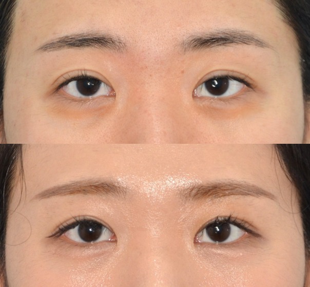 no.52 [Case Studies] Case 8. Lowering the Line with Double-Line Release + Ptosis Correction After double eyelid surgery, some people find their line too high, so it looks puffy like a sausage or makes them look sleepy, which causes stress and leads them to seek improvement. ▶ Double Eyelid Revision (2): Lowering the Line (Double-Line Release) You can read about the double-line release technique through the link above. Let's look at an actual case. This patient originally came in wanting lateral/lower canthoplasty. She was a candidate for lateral/lower canthoplasty, but she currently shows some sanpaku (scleral show below the iris), and rather than the closed-in look at the outer corners, improving the double eyelid shape and the slightly sleepy look would be far more effective for her eyes, so I recommended incisional ptosis correction. The line is high toward the inner side, and a fair amount of the dark iris is covered, showing mild ptosis. The plan was to lower the line with double-line release surgery and make the eyes more defined with ptosis correction. The new line was designed slightly lower than the old line toward the inner side, and after the incision, all adhesions of the old line were released. The eye-opening muscle was advanced 6 mm on both sides, performing ptosis correction by levator advancement, and the upper eyelid fat was used to cover the adhesion site of the old line, which completes the surgery. This is 2 months after surgery. The line has been lowered nicely, and the eyes look much more defined than before surgery while changing naturally. Double-line release (lowering the line) + ptosis correction: before / 2 months after Comparing before and after side by side makes the difference even clearer. This case shows well that a bigger double eyelid does not make the eyes look bigger. The patient was very satisfied, and I expect it to settle even more naturally with a little more time. ◀ Previous post: Case 7. Removing Multiple Double Eyelid Folds with Incisional Ptosis Correction Next post: Case 9. Partial Lateral Canthoplasty Reversal and Lower Canthoplasty Revision ▶ Related posts ▶ Double Eyelid Revision (1) ▶ Incisional Ptosis Correction: Technique, Principle, Pros and Cons ▶ Upper Eyelid Fat Repositioning: Effects and Surgical Method WhatsApp Inquiries https://api.whatsapp.com/message/DE3GWE4FQGMDF1?autoload=1&app_absent=0 Sai Plastic Surgery — Phone Inquiries Main Line: +82-2-512-4647 * This post was written by Dr. Kwang-rae Kang, Director of Sai Plastic Surgery, with his own text and illustrations, for the purpose of providing medical information in compliance with Article 56(1) of the Korean Medical Service Act. + Read more
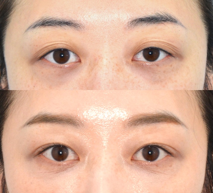 no.49 [Case Studies] Case 7. Removing Multiple Double Eyelid Folds with Incisional Ptosis Correction Some people find it uncomfortable to open their eyes because their double eyelid has multiple folds. I'd like to share a case where this was resolved. Analyzing the photo, there are multiple folds, and the eye on the right >> in the photo in particular looks sleepy. The patient also said that opening her eyes was difficult and uncomfortable, so she wanted the line neatly tidied up. Her eyes are slightly prominent, and slightly hollow above, an eye structure prone to developing multiple folds. The right eye also has weak eye-opening strength, so it looks as if it has even more folds. In cases like this, an incision is made to create a secure double eyelid, the adhesions underneath where extra folds tend to form are released, and the upper eyelid fat is brought down to prevent the folds from re-forming. Weak eye-opening strength is compensated for with ptosis correction. You can also see several creases in the closed-eye photo. The design involves excising part of the skin, as shown below. The orbital fat is found and repositioned to where the extra folds used to form, to prevent them from forming again. The surgery is completed with ptosis correction and double eyelid fixation. The ptosis correction was done a little more on the right to balance the two sides. The overall process is similar to revision surgery for lowering a double eyelid line. This is 1 week after surgery. There is relatively little swelling or bruising. She said opening her eyes had become easier from 1 week after surgery. This is 2 months after surgery. There is almost no swelling, and the line has come out nicely in a slim shape. If you compare before and after, you can see that the overall impression of the eyes has changed dramatically. When there are this many folds, an incision is likely to be needed. However, some cases can be treated without an incision, so please come in for a consultation and get an accurate diagnosis. Because the method is similar to surgery for lowering the line, the post below may be helpful. ▶ Double Eyelid Revision (2): Lowering the Line (Double-Line Release) ◀ Previous post: Case 6. Non-Incision Double Eyelid + Lateral/Lower Canthoplasty: Before/After (Asymmetry Correction) Next post: Case 8. Lowering the Line with Double-Line Release + Ptosis Correction ▶ Related posts ▶ Upper Eyelid Fat Repositioning: Effects and Surgical Method ▶ Incisional Ptosis Correction: Technique, Principle, Pros and Cons WhatsApp Inquiries https://api.whatsapp.com/message/DE3GWE4FQGMDF1?autoload=1&app_absent=0 Sai Plastic Surgery — Phone Inquiries Main Line: +82-2-512-4647 * This post was written by Dr. Kwang-rae Kang, Director of Sai Plastic Surgery, with his own text and illustrations, for the purpose of providing medical information in compliance with Article 56(1) of the Korean Medical Service Act. + Read more
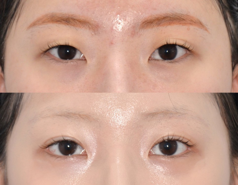 no.48 [Case Studies] Case 6. Non-Incision Double Eyelid + Lateral/Lower Canthoplasty: Before/After (Asymmetry Correction) This time, the case is a patient who wanted her asymmetry corrected. In the pre-operative photo, you can see that the << left eye is noticeably smaller than the right. It is more hooded above, and the area of white at the outer corner is also smaller than on the right. The >> right eye also protrudes a little more. The goal was to make the right eye even a little bigger to balance the two sides. For the double eyelid, the non-incision method was sufficient, so I recommended it, and I did not recommend epicanthoplasty because the caruncle (the pink tissue at the inner corner) was already well visible. For lateral and lower canthoplasty, we decided to open the left eye slightly more to balance the two sides. This is 4 months after surgery. Because the patient lives far from Seoul and could not visit the clinic often, she came in only for the minimum check-ups, so please understand that there are no progress photos. To make up for the smaller << left eye, the << left line was set with a slightly semi-out feel, and the >> right line as an in-out line. Surgery done asymmetrically like this is rare, so I chose this case to share. Lateral canthoplasty: << left 3 mm, >> right 2 mm Lower canthoplasty: << left 2 points, >> right 1 point so the surgery was done completely differently on each side. Before / 4 months after surgery If you compare before and after, you can see that the difference between the two eyes has been greatly reduced. Looking closely, a little asymmetry remains here and there, but considering that perfect symmetry is difficult to achieve, overall the eyes are much better balanced. As this case shows, correcting asymmetry sometimes requires doing the surgery asymmetrically, which can make it difficult. Please check carefully what plan the clinic proposes for correcting asymmetry before having surgery. Want to know about non-incision double eyelid surgery? ▶ Non-Incision Double Eyelid Surgery: Technique, Principle, Pros and Cons Want to know about lateral and lower canthoplasty? ▶ What Effect Does Dual Canthoplasty Have? ◀ Previous post: Case 5. Incisional Ptosis Correction + Epicanthoplasty: Before/After and Recovery Next post: Case 7. Removing Multiple Double Eyelid Folds with Incisional Ptosis Correction ▶ Related posts ▶ Types of Double Eyelid Lines ▶ Lower Canthoplasty: Method and Effects (Fascial Fixation vs. Periosteal Fixation) WhatsApp Inquiries https://api.whatsapp.com/message/DE3GWE4FQGMDF1?autoload=1&app_absent=0 Sai Plastic Surgery — Phone Inquiries Main Line: +82-2-512-4647 * This post was written by Dr. Kwang-rae Kang, Director of Sai Plastic Surgery, with his own text and illustrations, for the purpose of providing medical information in compliance with Article 56(1) of the Korean Medical Service Act. + Read more
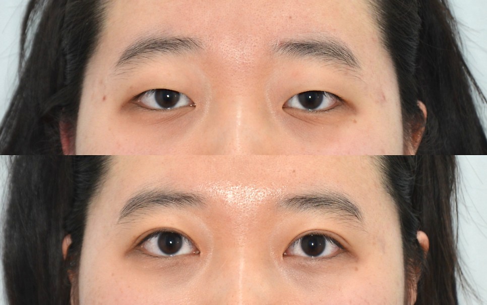 no.45 [Case Studies] Case 5. Incisional Ptosis Correction + Epicanthoplasty: Before/After and Recovery (feat. Scar Tissue) This time, let's look at a patient who had incisional ptosis correction together with epicanthoplasty, see what changed, and follow how the scars and swelling progressed. Before surgery, the dark part of the eye (the iris) was covered and less visible, giving the eyes a heavy, confined look, and you can see that she raised her eyebrows to open her eyes. She was also using the << (left in the photo) eyebrow more. A fairly prominent epicanthal fold is visible at the inner corners, along with a long crease extending downward. Analysis with AI software showed that about 75–80% of the iris was exposed. At least 85% is needed for the eyes not to look heavy, and around 90% exposure gives a clear, bright look. Her surgical plan was incisional double eyelid + ptosis correction and epicanthoplasty. This was because there was a lot of loose skin that needed to be removed, her eye-opening strength was weak, and the epicanthal fold at the inner corners was severe and needed improvement. This is the pre-operative design. About 2 mm of skin was excised on each side, and the epicanthoplasty was also about 2 mm on each side. For ptosis correction, on both sides equally, a 4.5 mm levator advancement was planned. 1 week after surgery: after stitch removal There was less swelling than expected, with slight bruising. The eyes look much clearer than before surgery, and you can see that the crease below was reduced by the epicanthoplasty. 1 month after surgery There was still some swelling, and in the left eye in the photo, at the inner and outer corners, the line appeared doubled. At the inner corner, this was due to slight scarring, so I gave a scar injection. The outer corner is something that gradually improves over time. This is 3 months after surgery. Almost all of the swelling has gone down, and the issues seen at 1 month have all improved. After surgery, iris exposure increased to over 90%, giving the eyes a clear, bright look. And the deep crease that started from the inner corner also improved with the epicanthoplasty. The epicanthoplasty scar is barely visible, and I expect it to improve further. This is how visible the incisional double eyelid scar is. It is still slightly visible at 3 months, but it will fade further and become invisible over time. Incisional double eyelid surgery and epicanthoplasty both involve incisions and sutures, so many people are afraid of scarring and of the recovery, which is why I wrote this post. Before / 3 months after Incisional ptosis correction / Epicanthoplasty Want to learn more about incisional ptosis correction? ▶ Incisional Ptosis Correction: Technique, Principle, Pros and Cons Want to learn more about epicanthoplasty? ▶ What Is Inner Corner Eye Surgery (Epicanthoplasty)? ◀ Previous post: Case 4. Non-Incision Double Eyelid Surgery: Before/After and Recovery Next post: Case 6. Non-Incision Double Eyelid + Lateral/Lower Canthoplasty: Before/After (Asymmetry Correction) ▶ Related posts ▶ How to Minimize Scarring After Double Eyelid or Epicanthoplasty Surgery ▶ Who Needs Ptosis Correction (Eye-Opening Surgery)? WhatsApp Inquiries https://api.whatsapp.com/message/DE3GWE4FQGMDF1?autoload=1&app_absent=0 Sai Plastic Surgery — Phone Inquiries Main Line: +82-2-512-4647 * This post was written by Dr. Kwang-rae Kang, Director of Sai Plastic Surgery, with his own text and illustrations, for the purpose of providing medical information in compliance with Article 56(1) of the Korean Medical Service Act. + Read more
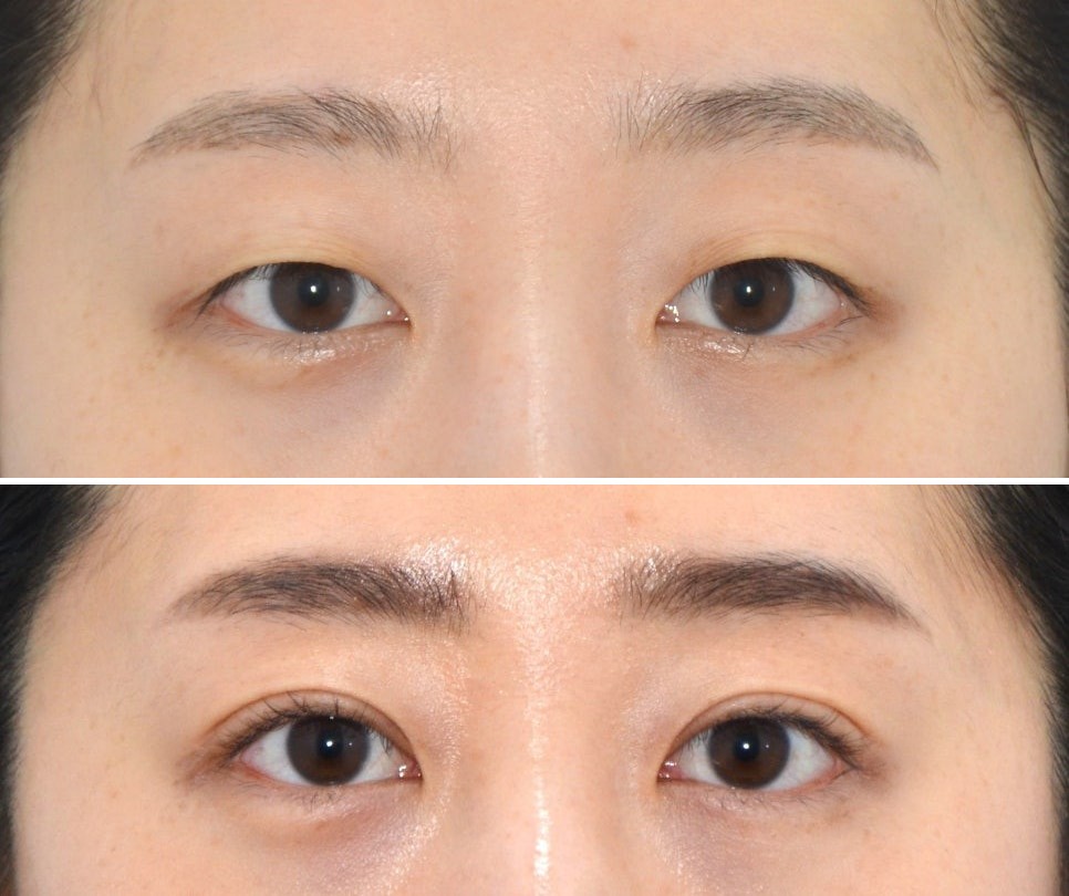 no.38 [Case Studies] Case 4. Non-Incision Double Eyelid Surgery: Before/After and Recovery Chief complaint She wanted an in-out double eyelid line and improvement of the multiple creases. Past history No previous surgery Assessment Her eyes are prominent (asymmetrically so; the eye on the right in the photo protrudes more), and her skin is on the thin side. Slight skin laxity creates some creases, and there is asymmetry in eye size. Surgical plan When I checked during the consultation, there was no difference in eye-opening strength, and the eyelids offered no resistance to forming a double eyelid, so I judged that the non-incision method would be sufficient. Actual surgery The structure required designing the inner part somewhat higher and the outer part slightly lower for an in-out line to form. The surgery was done with the non-incision method, at a height of 9 mm with 6 points. For non-incision surgery, we proceed without external stitches. Progress 1 week after surgery Because there are no stitches, we check the swelling and make sure there are no problems. There is swelling, and the tiny puncture marks on the skin are still visible. 1 month after surgery Much of the swelling has gone down, but a little remains, and the marks are still visible. The outer part of the line still looks slightly split. 3 months after surgery The swelling appears to have completely gone down, the look has become natural, and the marks have almost disappeared. As you can see, non-incision double eyelid surgery is not burdensome even during recovery, and its natural look is its strength. Curious about how non-incision double eyelid surgery is done? ▶ Non-Incision Double Eyelid Surgery: Technique, Principle, Pros and Cons ◀ Previous post: Case 3. Lower Eyelid Fat Repositioning (+ Removal): Before/After Next post: Case 5. Incisional Ptosis Correction + Epicanthoplasty: Before/After and Recovery ▶ Related posts ▶ Which Eyes Are Good Candidates for Non-Incision Double Eyelid Surgery? ▶ Types of Double Eyelid Lines WhatsApp Inquiries https://api.whatsapp.com/message/DE3GWE4FQGMDF1?autoload=1&app_absent=0 Sai Plastic Surgery — Phone Inquiries Main Line: +82-2-512-4647 * This post was written by Dr. Kwang-rae Kang, Director of Sai Plastic Surgery, with his own text and illustrations, for the purpose of providing medical information in compliance with Article 56(1) of the Korean Medical Service Act. + Read more
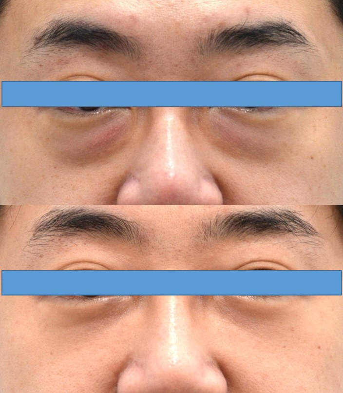 no.36 [Case Studies] Case 3. Lower Eyelid Fat Repositioning (+ Removal): Before/After Male, 40s Chief complaint Puffy bulges under the eyes. Past history No previous surgery Assessment From the front, bulging fat is visible, along with a hollow tear trough. The skin over the bulging area also looks reddish. The photo was taken with light shining only from above so that the overall shadowing is clearly visible. Along with the fat bulging forward, there is little fullness of the aegyo-sal (the roll under the eye), and the bulge extends a long way down. Surgical plan As anyone could tell, lower eyelid fat repositioning was needed, and since there was quite a lot of fat, fat removal was also required. Actual surgery Because there was too much fat, part of the fat was removed first, and then repositioning was performed. Repositioning alone, never removing any fat, is not always the answer. This is the fat that was removed. A similar amount was removed from each side. Because fat removal was needed, there was a little more bleeding, a bit more swelling, and the operation took slightly longer, so it was not an easy case. Even so, everything through the repositioning went perfectly. Progress Because there are no visible external stitches, we did not do a 1-week follow-up. 3 weeks after surgery The bulging fat under the eyes has decreased, and you can see that the redness of the skin has also improved. From the side, the height of the bulge has decreased, and the aegyo-sal is now much more visible. Because the light comes from above, there is slight shadowing below the aegyo-sal. Before / After He clearly looks much younger in person. In cases like this, if more time passes without surgery, the bulges become very prominent and make you look much older. I think it is better to have surgery early and enjoy a younger, cleaner-looking face. Curious about lower eyelid fat repositioning? ▶ Effects and Surgical Method of Lower Eyelid Fat Repositioning ◀ Previous post: Case 2. Lateral and Lower Canthoplasty + Lower Eyelid Fat Repositioning Next post: Case 4. Non-Incision Double Eyelid Surgery: Before/After and Recovery ▶ Related posts ▶ Effects and Surgical Method of Lower Eyelid Fat Repositioning ▶ Lower Blepharoplasty: Effects and Surgical Method (feat. Lower Eyelid Fat Repositioning) WhatsApp Inquiries https://api.whatsapp.com/message/DE3GWE4FQGMDF1?autoload=1&app_absent=0 Sai Plastic Surgery — Phone Inquiries Main Line: +82-2-512-4647 * This post was written by Dr. Kwang-rae Kang, Director of Sai Plastic Surgery, with his own text and illustrations, for the purpose of providing medical information in compliance with Article 56(1) of the Korean Medical Service Act. + Read more
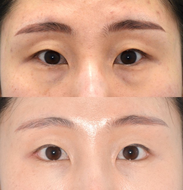 no.33 [Case Studies] Case 2. Lateral and Lower Canthoplasty + Lower Eyelid Fat Repositioning: Before/After and Recovery Female, early 30s Chief complaint She wanted lower eyelid fat repositioning and a natural-looking lateral and lower canthoplasty. She had already researched the surgeries she wanted and had made up her mind before coming in. Past history No previous surgery Front view before surgery Assessment Under-eye fat From the front, bulging fat is visible, along with a hollow tear trough. Along with the fat bulging forward, there is little fullness of the aegyo-sal (the roll under the eye), and you can see that the bulge of orbital fat extends a long way down. The area below the outer corner of the eye is heavily covered. Lateral and lower canthoplasty The outer corners of the eyes slant upward, giving the outer part of the eye a confined look. In the photo, the << left eye (the patient's actual right eye) is slightly higher. Measured with eye analysis software, the right eye is positioned 0.5 mm higher. The angle is also steep (the average is 4–8 degrees), and the right side is 2.5 degrees higher. The proportion of visible white on the outer side is about 20–21%. Surgical plan Just as the patient had thought, lower eyelid fat repositioning seemed appropriate, and additional procedures such as fat grafting did not seem necessary. For lateral canthoplasty, what matters is how much space there is beyond the outer corner, and fortunately this patient had plenty. Actual surgery Design This was a case where repositioning alone was sufficient, without removing any orbital fat. Lateral canthoplasty: 3 mm on each side. I usually open about 3 mm on average; at 3 mm, everyone notices that the eyes look longer. Lower canthoplasty: fixed at 2 points on each side. For lower canthoplasty I usually fix 2 points, and up to 3 points when needed. Progress 1 week after surgery The stitches were removed. There was a little chemosis (conjunctival swelling), slight swelling under the eyes, and some yellowish bruising. For 1 week, this is relatively little swelling and bruising. Even at this point, I thought the shape looked fine. The eye on the left side of the photo seemed to have a bit more chemosis. 1 month after surgery Lower eyelid fat repositioning The bulging and hollow areas under the eyes are barely visible. Because makeup makes it hard to see, looking from a 45-degree angle and from the side: The bulging area has gone in, and the hollow is no longer noticeable. Above all, from the side, the height of the bulge has decreased. Only about as much fullness as an aegyo-sal remains. The upward look at the outer corners has also improved. Lateral and lower canthoplasty From the front, you can see that the eyes look much bigger. A very slight chemosis is still visible. There is no squared-off deformity or pinched shape. The outer corners have been lowered by 1.2 mm and 1 mm respectively, and the angle has decreased by 3.7 degrees on each side. The area of visible white on the outer side is now about 25% on both sides, considerably wider than before surgery. The patient was satisfied, and so was I. She will come back at 3 months, and I expect no major change, just a slightly more natural look. ※ Patients who are happy with their results usually come in wearing makeup and nicely dressed up. P.S. These are selfies the patient sent me. The difference looks even bigger in her selfies than in the clinic photos, though I'm not sure why. Curious about lateral and lower canthoplasty? ▶ What Effect Does Dual Canthoplasty Have? Curious about lower eyelid fat repositioning? ▶ Effects and Surgical Method of Lower Eyelid Fat Repositioning ◀ Previous post: Case 1. Incisional Double Eyelid Revision and Epicanthoplasty After an Undone Incisional Double Eyelid Next post: Case 3. Lower Eyelid Fat Repositioning (+ Removal): Before/After ▶ Related posts ▶ Case 1. Incisional Double Eyelid Revision and Epicanthoplasty After an Undone Incisional Double Eyelid ▶ Does Lower Eyelid Fat Repositioning Undo Lateral and Lower Canthoplasty? WhatsApp Inquiries https://api.whatsapp.com/message/DE3GWE4FQGMDF1?autoload=1&app_absent=0 Sai Plastic Surgery — Phone Inquiries Main Line: +82-2-512-4647 * This post was written by Dr. Kwang-rae Kang, Director of Sai Plastic Surgery, with his own text and illustrations, for the purpose of providing medical information in compliance with Article 56(1) of the Korean Medical Service Act. + Read more
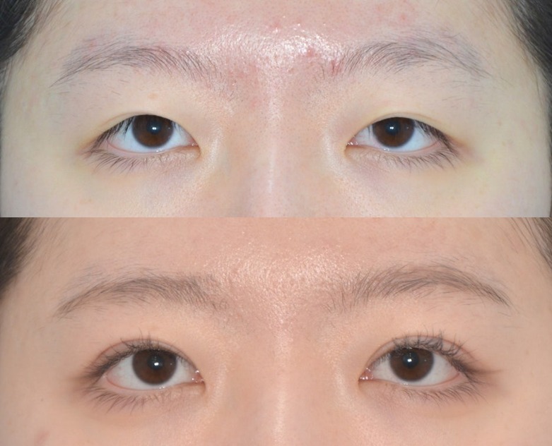 no.31 [Case Studies] Case 1. Incisional Double Eyelid Revision and Epicanthoplasty After an Undone Incisional Double Eyelid: Before/After and Recovery Female, early 20s Chief complaint "My double eyelid has come undone." "I look sleepy and tired." "I'd like the line a little higher." "Is it possible without an incision?" Past history Incisional ptosis correction 4 years ago. Before surgery Assessment The line had almost completely come undone, and the skin was covering the dark part of the eye (the iris). The right side in the photo (the patient's actual left eye) looked worse in particular. Her eye-opening strength was actually fine. Because the double eyelid had come undone, the skin had drooped down and covered the eye, which appeared to be a kind of pseudoptosis. Surgical plan I recommended incisional revision surgery. Because her eye-opening strength was fine, I did not recommend ptosis correction. The distance between her eyes was slightly wide, so to slightly improve the epicanthal fold and to make the inner part of the double eyelid line a little more visible, I recommended a small epicanthoplasty. Actual surgery Design To improve the existing incision scar, the line was placed just below the scar, 1.5 mm of skin was excised above it, and the double eyelid height was designed at 8 mm with the skin stretched (pulled taut). There was a lot of scar tissue under the skin from the previous surgery, and since her double eyelid had come undone despite incisional surgery, sufficient pretarsal tissue was removed to create a firmer adhesion. The epicanthoplasty was performed with the Hiraga method, 1 mm on each side. Progress 1 week after surgery The stitches were removed, and there was a fair amount of swelling. Fortunately, there was almost no bruising. Perhaps because of the swelling, the eyes looked slightly sleepy. Because the epicanthoplasty was small, the surgical marks were not noticeable. 1 month after surgery There was a bit more swelling than usual for one month, so I recommended anti-swelling injections. 2 months after surgery She said the swelling went down quickly after the anti-swelling injections, and her eyes now look clearer than before. An eyelash perm is much more effective when you have a double eyelid. It is not perfectly symmetrical like a mirror image, but the patient was satisfied, and so was I. I hope it never comes undone again. ※ Patients who are happy with their results usually come in wearing makeup and nicely dressed up. Next post: Case 2. Lateral and Lower Canthoplasty + Lower Eyelid Fat Repositioning ▶ Related posts ▶ Double Eyelid Revision (1): Raising the Line ▶ Incisional Double Eyelid Surgery: How Is It Done? WhatsApp Inquiries https://api.whatsapp.com/message/DE3GWE4FQGMDF1?autoload=1&app_absent=0 Sai Plastic Surgery — Phone Inquiries Main Line: +82-2-512-4647 * This post was written by Dr. Kwang-rae Kang, Director of Sai Plastic Surgery, with his own text and illustrations, for the purpose of providing medical information in compliance with Article 56(1) of the Korean Medical Service Act. + Read more